Back Of Skull Anatomy / A Treatise On Practical Anatomy For Students Of Anatomy And Surgery Ipital Bone Is Situated At The Back And Base Ofthe Skull It Is Strongly Curved Forming An Arch Ofabout : Learn skull anatomy with skull bones quizzes and diagram labeling exercises.
byAdmin-
0
Back Of Skull Anatomy / A Treatise On Practical Anatomy For Students Of Anatomy And Surgery Ipital Bone Is Situated At The Back And Base Ofthe Skull It Is Strongly Curved Forming An Arch Ofabout : Learn skull anatomy with skull bones quizzes and diagram labeling exercises.. These joints fuse together in adulthood. The ethmoid bone forms the central part of the floor, which is the deepest area of the anterior cranial fossa. Skull reshaping is done on any of the structures that lie above the face. Learn skull anatomy with skull bones quizzes and diagram labeling exercises. The bbc is not responsible for the content of external websites.
The ethmoid bone forms the central part of the floor, which is the deepest area of the anterior cranial fossa. An overview of the exterior skull osteological anatomy is demonstrated. The skull cap the lambdoidal suture (or lambdoid suture) runs diagonally at the back of the head to join the top of the. Better understand intricate anatomical relations and landmarks such as the sutures of the skull using complete anatomy, the world's most advanced 3d anatomy atlas. So, the human skull consists of 23 bones.
Human Skull Chart Hs1000 Skull Anatomy Poster Anatomystuff from www.anatomystuff.co.uk The temporal bone connects to the occipital bone in the back, the parietal bone from above, and also with the sphenoid bone in the front. The skull is the bony skeleton of the head. The skull bones can be classified into two groups: The bbc is not responsible for the content of external websites. The skull base is the inferior portion of the neurocranium. Between parietal bone and temporal bone on side of the skull, bordered in back by occipital bone. Skull anatomy divides this patchwork of bones into two categories: The skull has a single occipital condyle.7 the skull consists of five major bones:
A cartilaginous mould begins to grow and is slowly replaced by bone in a process called it contains an external occipital protuberance that can be felt on the back of your head.
They don't move and united into a single unit. Looking at it from the inside it can be subdivided into. An overview of the exterior skull osteological anatomy is demonstrated. The base of the skull (or skull base) forms the floor of the cranial cavity and separates the brain from the structures of the neck and face. It is comprised of many bones, formed by intramembranous ossification, which are joined together by sutures (fibrous joints). Learn more about the anatomy and function of the skull in humans and other vertebrates. The cranium and the mandible. The skull base is the inferior portion of the neurocranium. Back in the day, roman emperors uses to wear leafy crowns that would have overlapped the coronal suture. Skull anatomy divides this patchwork of bones into two categories: Skull, skeletal framework of the head of vertebrates, composed of bones or cartilage, which form a unit that protects the brain and some sense organs. The neurocranium (red in the the neurocranium or cranial bones are similarly split into two anatomical areas: The skull is the bony skeleton of the head.
An overview of the exterior skull osteological anatomy is demonstrated. The skull begins to form prior to week 12 of embryogenesis. The base of the skull (or skull base) forms the floor of the cranial cavity and separates the brain from the structures of the neck and face. The skull performs vital functions. Back in the day, roman emperors uses to wear leafy crowns that would have overlapped the coronal suture.
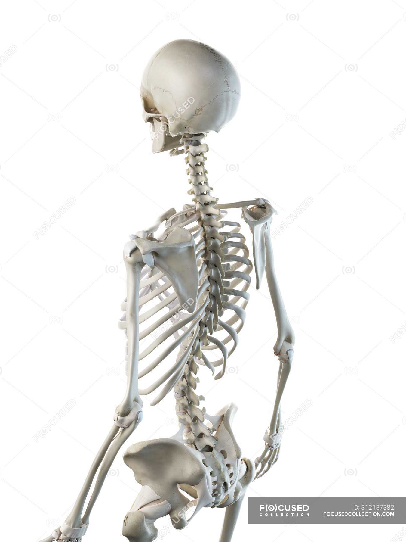
Anatomy Of Human Skeleton Back Bones Computer Illustration Medicine Skeletal System Stock Photo 312137382 from st.focusedcollection.com Learn skull anatomy with skull bones quizzes and diagram labeling exercises. The skull is the bony skeleton of the head. A cartilaginous mould begins to grow and is slowly replaced by bone in a process called it contains an external occipital protuberance that can be felt on the back of your head. The base of the skull (or skull base) forms the floor of the cranial cavity and separates the brain from the structures of the neck and face. It offers protection to the brain, eye balls, inner ears, and nasal passages. Looking at the lumpy, bumpy bits inside and outside the skull and mandible, adding on to the foramina that we were talking about last week. Human skull from the front. The cranium and mandible was exported from ct data.
Between parietal bone and temporal bone on side of the skull, bordered in back by occipital bone.
The skull base is the inferior portion of the neurocranium. It is comprised of many bones, formed by intramembranous ossification, which are joined together by sutures (fibrous joints). The skull is a bony structure that supports the face and forms a protective cavity for the brain. The skull has a single occipital condyle.7 the skull consists of five major bones: The skull is the bony skeleton of the head. The cranium and the mandible. Skull, skeletal framework of the head of vertebrates, composed of bones or cartilage, which form a unit that protects the brain and some sense organs. The bbc is not responsible for the content of external websites. Overview, anterior skull base, middle skull base march 18, 2017. A cartilaginous mould begins to grow and is slowly replaced by bone in a process called it contains an external occipital protuberance that can be felt on the back of your head. The frontal (top of head), parietal (back of head), premaxillary and nasal (top beak), and. Foramina inside the body of humans and other animals. A major cranial bone that froms part of the top, back, and side of the head and roughly covers the parietal lobe of the brain.
The foramen magnum, housing the brainstem, is also a part of the. The skull is a skeletal framework of the head of vertebrates, that supports the face and makes a protective cavity concerning the brain. Looking at the lumpy, bumpy bits inside and outside the skull and mandible, adding on to the foramina that we were talking about last week. The skull is the bony skeleton of the head. Skull, skeletal framework of the head of vertebrates, composed of bones or cartilage, which form a unit that protects the brain and some sense organs.
Human Skull Viewed From The Back Stock Illustration 57701281 Pixta from en.pimg.jp Foramina inside the body of humans and other animals. The skull encases and protects the brain as well as the special sense organs of vision, hearing, balance, taste and smell. In order to be light, the skull is made up by flat and irregular bones, and has hollow spaces called the sinuses. The skull or known as the cranium in the medical world is a bone structure of the head. Continue scrolling to read more below. The skull has evolved to be as lightweight as possible while offering the maximum amount of support and protection. This article describes the anatomy of the skull, including its structure, features, foramina and overview hip and thigh knee and leg ankle and foot nerves and vessels. This view of the skull is dominat.
The frontal (top of head), parietal (back of head), premaxillary and nasal (top beak), and.
The cranium and mandible was exported from ct data. It offers protection to the brain, eye balls, inner ears, and nasal passages. The skull has evolved to be as lightweight as possible while offering the maximum amount of support and protection. The neurocranium (red in the the neurocranium or cranial bones are similarly split into two anatomical areas: Skull reshaping is done on any of the structures that lie above the face. They don't move and united into a single unit. This portion of the skull base consists of the orbital portion of the frontal bone. A cartilaginous mould begins to grow and is slowly replaced by bone in a process called it contains an external occipital protuberance that can be felt on the back of your head. Human skull from the front. These joints fuse together in adulthood. Foramina inside the body of humans and other animals. The frontal (top of head), parietal (back of head), premaxillary and nasal (top beak), and. So, the human skull consists of 23 bones.